Scientists Were Surprised By What The Human Eye Does After Death
There are a few standout elements to Frankenstein's monster that rely heavily on the fiction side of science fiction. One of those being that Frankenstein's monster possesses a revived brain and is perfectly able to use said brain in interpreting visual cues. See, even if Dr. Frankenstein had found a beautifully preserved brain and wonderfully intact eyes, it would have been impossible to bring those dead parts back to life. Or so it was commonly thought. More recently, researchers have been able to facilitate light signaling in the postmortem retinas. Is this the start of reviving dead nervous tissue?
A paper published in Nature in 2022, displayed a fascinating challenge to the commonly established theory that the death of brain cells is completely irreversible. The retina is a unique outgrowth of the central nervous system, composed of rods and cones which translate light information into the brain's language: electricity. Examining human eyes provided through organ donation, the authors found that these photoreceptors continued to respond to light for as long as five hours after the patient had passed.
Connecting the circuit: how other cells in the visual process might be preserved
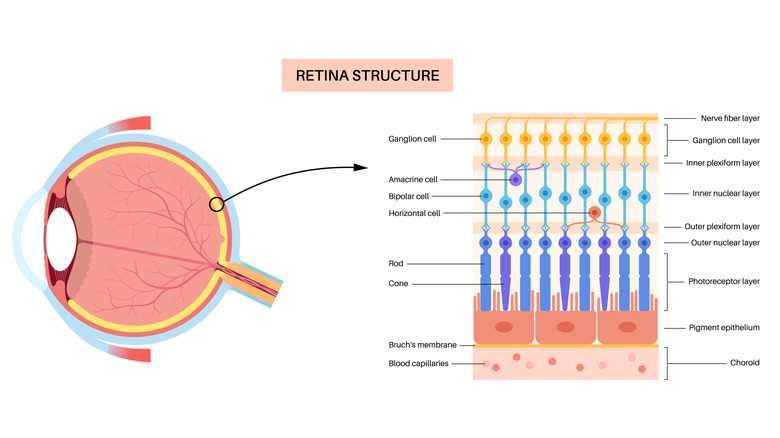
Although this in itself was fascinating, the authors noticed that these photoreceptors were not effectively transmitting this information to bipolar cells. These cells essentially interpret information from the photoreceptors in the outer retina and translate it for cells in the inner retina, acting as vital intermediaries in conveying visual cues. So, these researchers examined the activity of retinal cells in mice via electroretinogram (ERG) immediately following death . They found that photoreceptors continued emitting activity far longer than bipolar cells.
The next question was why are these bipolar cells losing all activity so quickly after death? To investigate this, the authors altered the environment of these cells to mimic certain typical physiological elements. They found that deoxygenation was a rapid driver of bipolar cell inactivity. Thus, they designed a system that could maintain oxygenation while human organ donor eyes were transported. Using this system, they were able to record electrical activity through ERG that was indicative of bipolar cell activation. This marked the first time that such activity had been recorded from the postmortem human retina.
Looking to the future of research

Given that these photoreceptors are specialized sensory neurons, the authors approached the retina as a model for the greater central nervous system, suggesting that similar techniques could be implemented to investigate postmortem neuronal activity elsewhere. However, while this is very promising and innovative work, there are still many steps before these active cells can effectively communicate among each other and lead to more ambitious innovations.
Though Mary Shelley's literary nightmares remain far from feasible, the future use of these techniques could really open up research possibilities. For instance, this model might allow scientists to study other nervous tissue in the context of neurodegenerative diseases like Alzheimer's disease. Additionally, if retinal cells can have a longer functional life past death, this could mean great things for future transplant of healthy retinal cells in cases of visual impairment, such as macular degeneration. Although, how the integration of these cells could be effective in living retinal circuits remains unknown. The authors, however, suggest that the electrical activity that they observed could be used to mark effective surgically transplanted retina.